



Digital X-Ray (RVG) in Patna for Clearer Dental Diagnosis
Dental Comfort Zone, Patna uses digital dental X-ray technology, commonly referred to as RVG (RadioVisioGraphy), to capture dental images quickly and display them digitally for clinical assessment.
Digital radiography can support dentists in evaluating teeth and surrounding structures for selected dental conditions, treatment planning and follow-up care. Your dentist decides when an X-ray is clinically appropriate based on your individual needs.
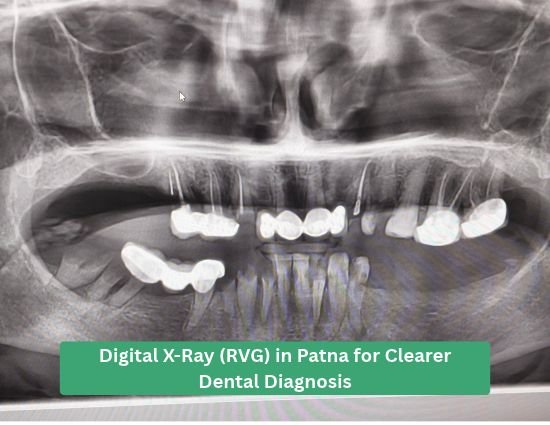
What Is Digital X-Ray (RVG)?
RVG stands for RadioVisioGraphy and refers to a digital dental radiography system in which an electronic sensor captures an X-ray image and makes it available digitally.
Compared with traditional film-based dental radiography, digital imaging can make image viewing and clinical communication more convenient because the captured image can be displayed on a computer or digital screen.
At Dental Comfort Zone, digital X-rays may be recommended when the dentist needs additional information that cannot be obtained from a routine clinical examination alone.
Digital Image Capture
Dental X-ray images are captured using a digital sensor and displayed electronically for clinical assessment.
Quick Image Viewing
Digital images can generally be viewed soon after acquisition, supporting efficient clinical evaluation.
Easy Digital Storage
Digital radiographic images can be stored electronically as part of the patient's clinical records.
Treatment Support
Radiographic findings can provide additional information to support diagnosis and treatment planning when clinically indicated.
Why Dental Clinics Use Digital X-Ray (RVG)
Digital radiography can make dental imaging more convenient for clinicians and patients while providing useful diagnostic information when an X-ray is clinically justified.
Fast Digital Image Availability
Digital images can generally be viewed soon after acquisition, helping the dentist assess the relevant area during the consultation.
Detailed Dental Imaging
Dental X-rays can reveal information about teeth and supporting structures that may not be visible during a routine visual examination.
Easy Image Sharing
Digital images can be displayed on a screen and may be easier to incorporate into digital clinical communication and records.
Supports Treatment Planning
When indicated, radiographic findings can help the dental team plan procedures and evaluate specific dental concerns.
Digital Record Keeping
Digital radiographs can be stored electronically and reviewed later as part of appropriate clinical documentation.
Patient-Friendly Explanation
Displaying the dental image on a screen can help dentists explain clinical findings and treatment considerations to patients.
Experienced Doctors Using Digital X-Ray for Precise Dental Care
At Dental Comfort Zone, our dentists combine clinical expertise with digital dental imaging to make diagnosis and treatment planning more accurate, efficient, and patient-focused.

Dr. Rohit Singh
BDS, MDS/ODS, MS, FICOI (USA), PhD Scholar
Dr. Rohit Singh uses digital dental X-rays as an important diagnostic tool during implant and complex dental treatment planning. RVG images help him assess tooth and root structures, evaluate surrounding bone and identify dental conditions that require closer clinical evaluation.
How RVG Supports His Treatment
- Initial assessment of teeth and surrounding structures
- Root and bone evaluation before dental procedures
- Support for implant and restorative treatment planning
- Assessment of complex dental conditions
- Monitoring treatment progress where appropriate

Dr. Neha Singh
BDS, MDS
Dr. Neha Singh uses digital X-ray imaging to support diagnosis in restorative, cosmetic and endodontic treatments. RVG can provide a quick view of teeth and root structures, helping her evaluate dental concerns and plan appropriate treatment before and during procedures.
How RVG Supports Her Treatment
- Assessment of tooth and root conditions
- Support for root canal and endodontic diagnosis
- Evaluation before restorative and cosmetic procedures
- Identifying areas requiring further clinical assessment
- Supporting treatment planning and follow-up
When Can a Digital Dental X-Ray Be Useful?
The dentist determines whether an X-ray is necessary based on the patient's symptoms, examination findings and treatment requirements.
Root Canal Assessment
Dental radiographs can provide important information about the tooth and surrounding structures during selected root canal assessments and treatment planning.
- Assessment of tooth structure
- Evaluation around the root
- Treatment planning support
Dental Pain & Diagnosis
When clinically appropriate, radiographic imaging may help the dentist investigate certain causes of dental pain or other symptoms.
- Additional diagnostic information
- Assessment of selected dental conditions
- Support for clinical decision-making
Dental Treatment Planning
X-ray findings can contribute to treatment planning when the dentist needs information about structures that cannot be assessed clinically.
- Pre-treatment assessment
- Evaluation of selected teeth
- Treatment planning support
Dental Implants
Conventional dental X-rays may be used in selected stages of implant assessment, while more detailed 3D imaging such as CBCT may be required when indicated.
- Selected implant assessments
- Supporting clinical evaluation
- CBCT when 3D information is required
Impacted or Developing Teeth
Radiographic imaging may help dentists evaluate the position and development of selected teeth when clinically required.
- Tooth position assessment
- Supporting orthodontic evaluation
- Additional diagnostic information
Follow-Up Evaluation
When clinically justified, digital radiographs may be used to compare findings over time or assess selected treatment outcomes.
- Follow-up imaging
- Comparison with previous images
- Ongoing clinical assessment
Digital X-Ray (RVG) vs Traditional Film X-Ray
Digital radiography changes how dental X-ray images are captured, viewed and managed compared with traditional film-based radiography.
| Feature | Digital X-Ray (RVG) | Traditional Film |
|---|---|---|
| Image capture | Digital sensor | Conventional dental film |
| Image viewing | Digital display | Film processing required |
| Digital storage | Can be stored electronically | Physical film storage |
| Clinical communication | Easy digital viewing and display | Physical film handling |
| Processing workflow | Digital workflow | Chemical film processing |
Actual image quality and radiation exposure depend on the imaging system, sensor, exposure settings, positioning and examination protocol. Dental X-rays should be prescribed when clinically justified.
Your Digital X-Ray Experience
A digital dental X-ray is generally a quick imaging procedure. Your dentist will explain why the image is needed and what area will be examined.
Clinical Examination
Your dentist first examines your teeth and discusses your symptoms or treatment requirements.
X-Ray Recommendation
If additional information is required, your dentist may recommend an appropriate dental X-ray.
Digital Image Capture
The digital sensor is positioned for the required area and the X-ray image is captured using the appropriate imaging protocol.
Image Review
The captured image can be displayed digitally so your dentist can evaluate the relevant dental structures.
Diagnosis & Treatment Planning
Your dentist combines the radiographic findings with your clinical examination and other relevant information before recommending treatment.
Dental X-Rays Should Be Used When Clinically Needed
Dental X-rays use ionising radiation, so radiographic examinations should be recommended when the expected clinical benefit justifies the exposure.
The radiation dose from an individual examination depends on factors such as the equipment, exposure settings, imaging area and examination protocol.
- X-rays are recommended based on clinical need.
- The required imaging area is selected according to the diagnostic question.
- Appropriate exposure settings are used for the examination.
- Digital imaging supports efficient image review and electronic record keeping.
- Your dentist decides whether conventional digital X-ray or another imaging modality is more suitable.
Digital X-Ray or CBCT — Which One Is Needed?
Digital X-ray and CBCT are different dental imaging technologies. They should not be treated as interchangeable examinations.
A conventional digital dental X-ray may be sufficient when the dentist needs a focused two-dimensional image of a particular area. CBCT may be considered when three-dimensional information is clinically necessary.
The choice depends on the diagnostic question, treatment requirements and individual patient assessment.
Your dentist may choose:
- Digital X-Ray (RVG) for selected focused dental imaging needs.
- CBCT when three-dimensional information is required.
- Both when different imaging information is clinically justified.
- Neither when imaging is not necessary for the clinical situation.

Technology-Assisted Dental Diagnosis
Dental Comfort Zone is a multi-specialty dental clinic in Patna, established in 2014 by Dr. Rohit Singh and Dr. Neha Singh.
The clinic combines experienced dental specialists, modern diagnostic technology and patient-focused treatment planning for routine and complex dental care.
Diagnostic imaging is selected according to individual clinical requirements. Your dentist will explain why an X-ray may be recommended and how the findings may contribute to your treatment plan.
Digital X-Ray (RVG) FAQs
Get the Right Dental Imaging for Your Treatment
Whether you need a routine dental assessment, root canal evaluation, treatment planning or another dental procedure, our team can determine whether digital X-ray or another imaging technology is appropriate for your case.